
Identifying x rays haspi answer key – Exploring the realm of identifying X-rays for HASPI (Healthcare Associated Secondary Placental Insufficiency) diagnosis, this guide unveils the significance of X-rays in detecting this critical maternal and fetal health condition. Understanding the characteristic X-ray findings associated with HASPI empowers healthcare professionals to make accurate and timely diagnoses, leading to improved patient outcomes.
Delving into the intricacies of HASPI, this guide examines its causes, risk factors, symptoms, and potential complications. By recognizing the subtle signs and patterns visible on X-rays, clinicians can differentiate HASPI from other conditions with similar presentations, ensuring appropriate management strategies.
Identifying X-rays
X-rays are a form of electromagnetic radiation with high energy and short wavelength, commonly used in medical imaging. They are particularly useful for examining dense tissues such as bones, and are widely employed in various medical fields for diagnostic purposes.X-rays
are produced by passing a high-voltage electric current through a vacuum tube, causing electrons to strike a metal target. This collision generates X-rays, which can then penetrate the body and interact with different tissues. Denser tissues, such as bones, absorb more X-rays, resulting in a higher density on the resulting image.
This allows medical professionals to visualize and assess the internal structures of the body, aiding in the diagnosis of various medical conditions.There are different types of X-rays, each with specific applications. Plain X-rays, also known as conventional X-rays, are the most common type and provide a general overview of the targeted area.
Contrast X-rays, on the other hand, involve the administration of a contrast agent, such as barium or iodine, to enhance the visibility of certain organs or structures. Specialized X-ray techniques, such as fluoroscopy and computed tomography (CT), offer dynamic imaging and cross-sectional views, respectively.The
Healthcare Associated Secondary Placental Insufficiency (HASPI) score is a tool used to identify and assess the risk of HASPI, a condition that affects placental function and can lead to adverse pregnancy outcomes. HASPI is characterized by inadequate blood flow to the placenta, resulting in impaired oxygen and nutrient delivery to the developing fetus.HASPI
can be caused by various factors, including maternal hypertension, preeclampsia, and placental abruption. It is associated with an increased risk of intrauterine growth restriction, preterm birth, and stillbirth. Early identification and management of HASPI are crucial for improving pregnancy outcomes.
HASPI (Healthcare Associated Secondary Placental Insufficiency)
HASPI, or Healthcare Associated Secondary Placental Insufficiency, is a condition that affects the placenta, the organ responsible for providing oxygen and nutrients to the developing fetus during pregnancy. HASPI occurs when the placenta is unable to function properly, leading to inadequate blood flow and oxygen delivery to the fetus.The
causes of HASPI are not fully understood, but it is believed to be associated with certain maternal conditions, such as preeclampsia, hypertension, and placental abruption. Risk factors for HASPI include advanced maternal age, obesity, and a history of previous placental problems.Symptoms
of HASPI can include reduced fetal growth, decreased fetal movement, and abnormal uterine blood flow patterns. Complications of HASPI can be severe, including intrauterine growth restriction, preterm birth, and stillbirth.
HASPI and X-ray Identification
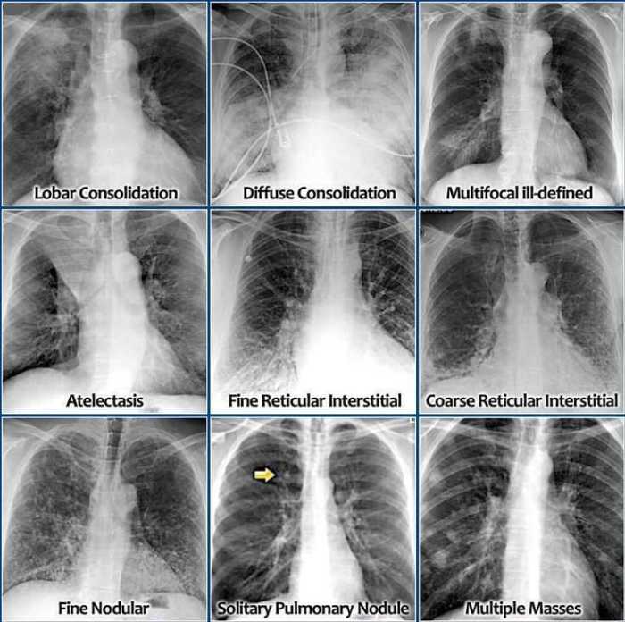
X-rays play a crucial role in the diagnosis of HASPI. By examining the placenta and surrounding structures, X-rays can reveal signs and symptoms that may indicate HASPI.One of the most common X-ray findings in HASPI is placental enlargement. This occurs due to the compensatory growth of the placenta in an attempt to increase the surface area for oxygen and nutrient exchange.
Other X-ray findings that may suggest HASPI include placental calcifications, which appear as white or dense areas on the X-ray image, and placental abruption, which is characterized by the separation of the placenta from the uterine wall.X-rays are a valuable tool for identifying HASPI, but it is important to note that other conditions can also cause similar X-ray findings.
Therefore, a thorough evaluation of the patient’s medical history, clinical presentation, and other diagnostic tests is necessary to confirm the diagnosis of HASPI.
Differential Diagnosis of HASPI
Differential diagnosis is crucial in identifying HASPI, as other conditions can present with similar X-ray findings. These conditions include:
Placental abruption
This condition occurs when the placenta separates from the uterine wall before the baby is born. It can cause sudden and severe abdominal pain and vaginal bleeding.
Placental previa
This condition occurs when the placenta implants in the lower part of the uterus, covering the cervix. It can cause painless vaginal bleeding during the second or third trimester of pregnancy.
Uterine fibroids
These are noncancerous growths in the muscular wall of the uterus. They can cause pelvic pain, heavy menstrual bleeding, and infertility.
Polyhydramnios
This condition occurs when there is too much amniotic fluid surrounding the baby. It can cause abdominal pain, swelling, and difficulty breathing.
Oligohydramnios
This condition occurs when there is too little amniotic fluid surrounding the baby. It can cause fetal growth restriction and other complications.To differentiate between HASPI and other conditions, doctors will consider the patient’s medical history, clinical presentation, and other diagnostic tests, such as ultrasound and blood tests.
Management of HASPI: Identifying X Rays Haspi Answer Key
The management of HASPI depends on the severity of the condition and the gestational age of the fetus. In mild cases, conservative management may be sufficient. This may include bed rest, close monitoring of the fetus, and medication to improve blood flow to the placenta.In
more severe cases, surgical intervention may be necessary. This may involve delivering the baby early or performing a cesarean section. The goal of treatment is to improve oxygen and nutrient delivery to the fetus and prevent adverse pregnancy outcomes.
FAQ Insights
What is the significance of X-rays in HASPI diagnosis?
X-rays provide valuable insights into the structural and functional abnormalities of the placenta, allowing clinicians to identify characteristic findings suggestive of HASPI.
How can X-rays differentiate HASPI from other conditions with similar presentations?
By analyzing specific X-ray findings, such as placental enlargement, calcifications, and abnormal vascular patterns, clinicians can distinguish HASPI from conditions like preeclampsia and placental abruption.
What are the key X-ray findings associated with HASPI?
Enlarged placenta, increased placental echogenicity, placental calcifications, and abnormal placental vascularity are among the characteristic X-ray findings indicative of HASPI.
